- Student
- Student
- News
- Events
- Business School
- Doctoral School
- International Dean’s Office
- International Cooperation Office
- Academic Career Office
- Academic Sports Association
- Library
- Student Groups
- Internships
- Electronic Examination Center
- Scholarship office
- Finance Department
- Your Stay in Poland
- Academic Schedule
- The Centre for People with Special Needs
- Alumni and Students Association
- Personal Student Profile
- Support Zone
- Virtual University
- Contact
- Individual Study Plan
- Empty
- Admissions
- Research
- University
- Erasmus+
Field of study
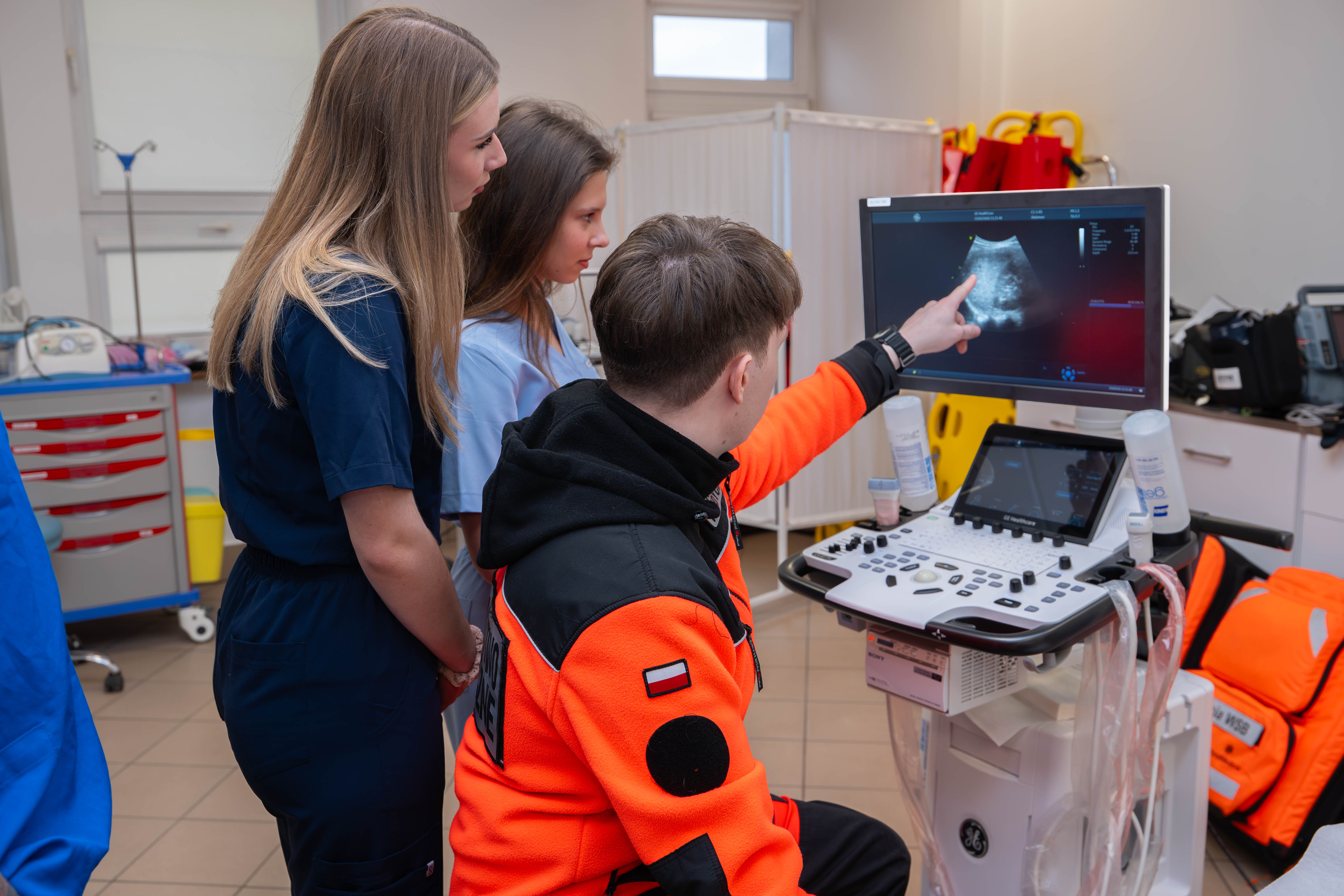
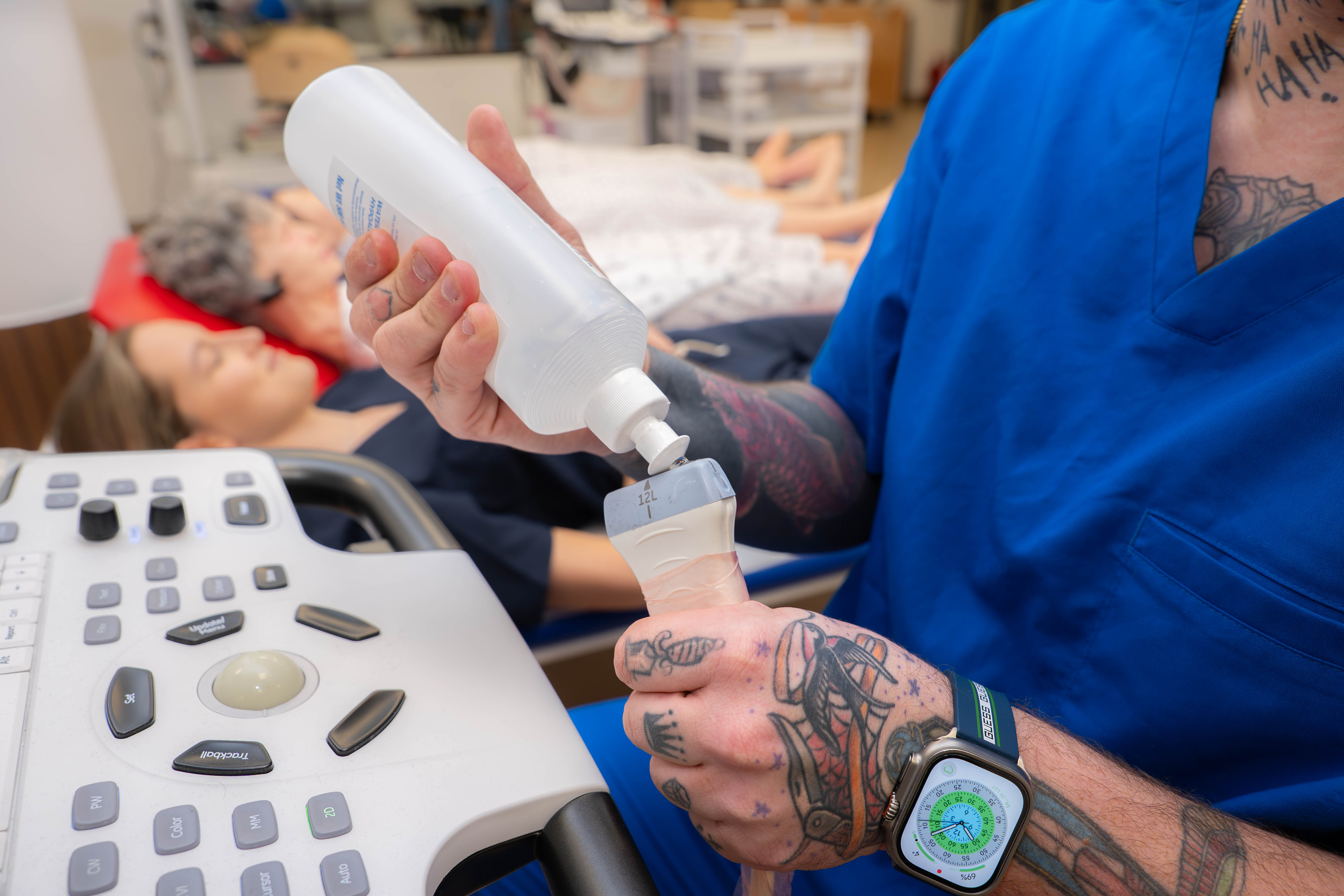
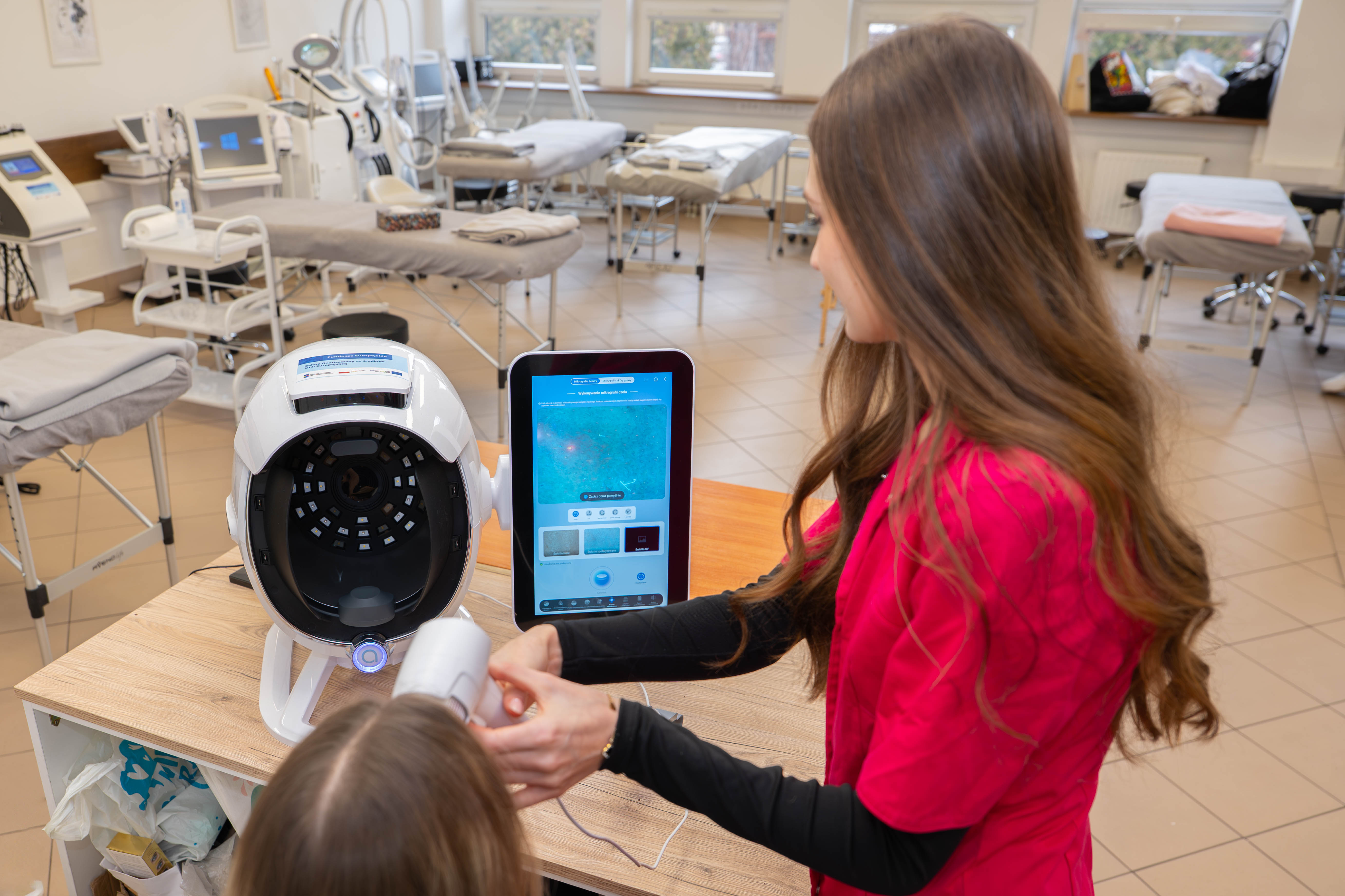
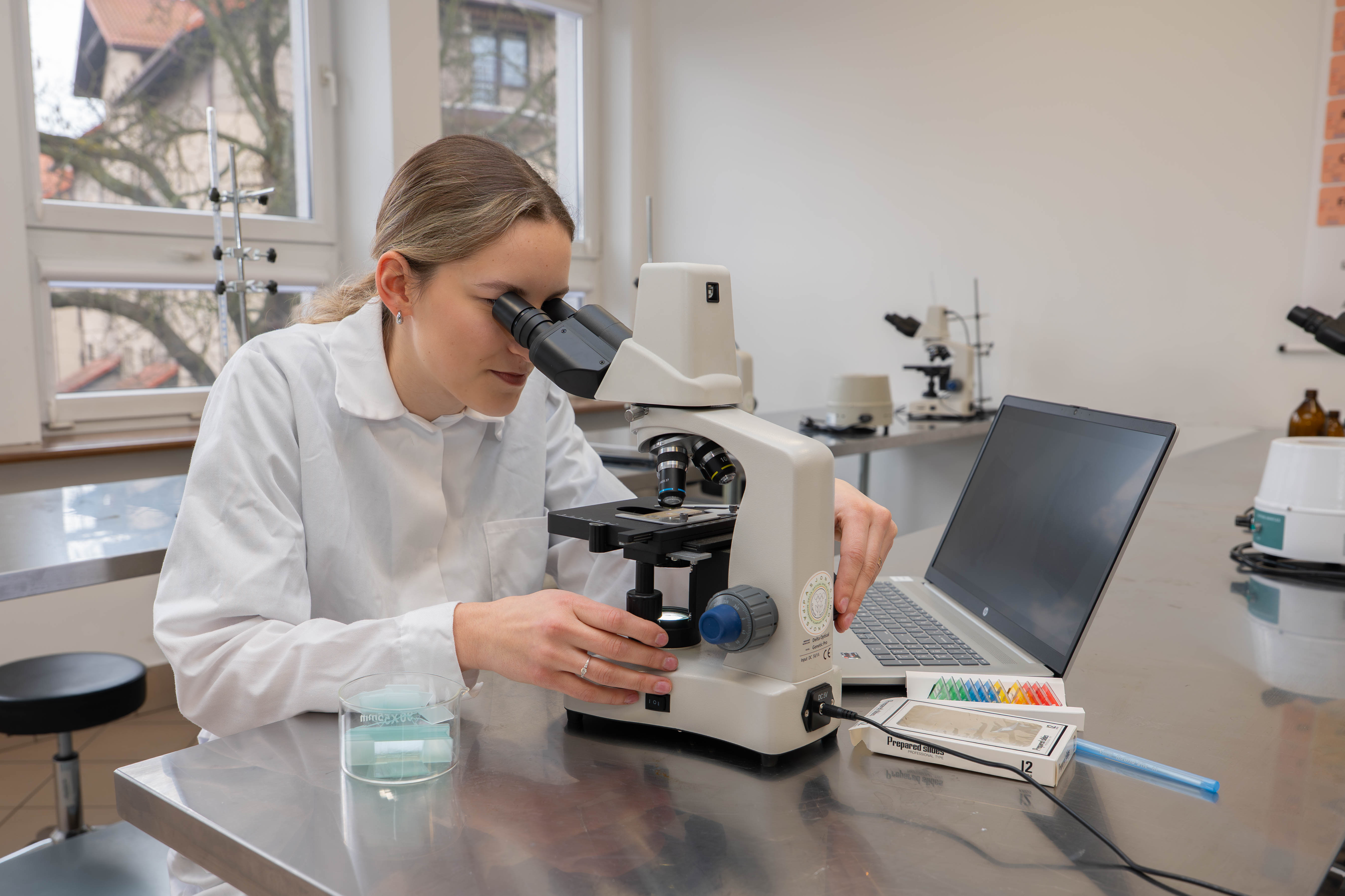
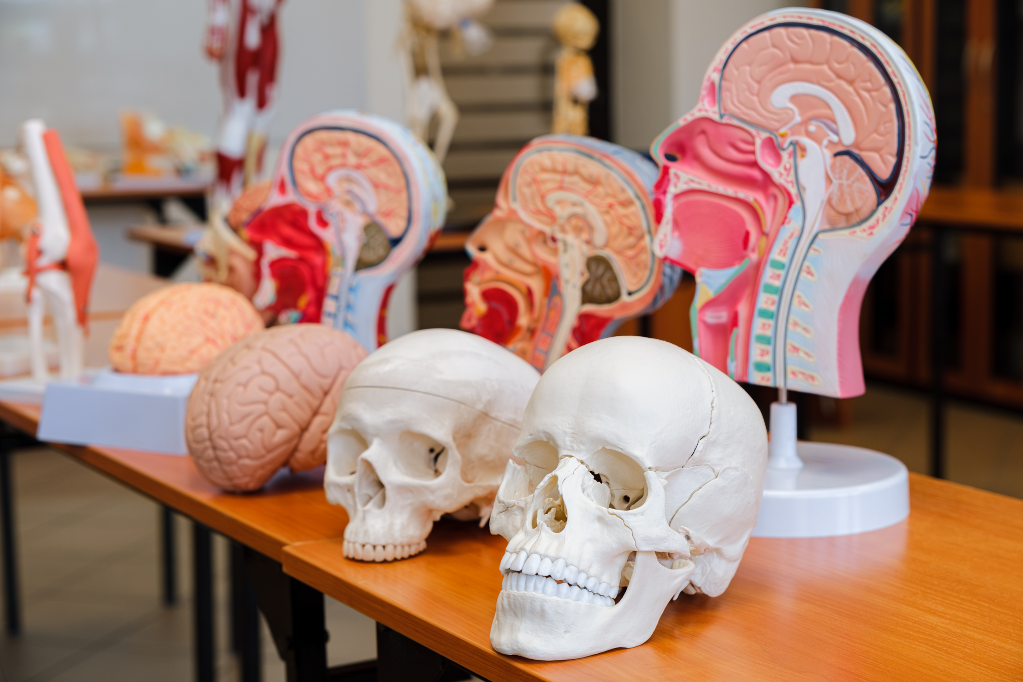
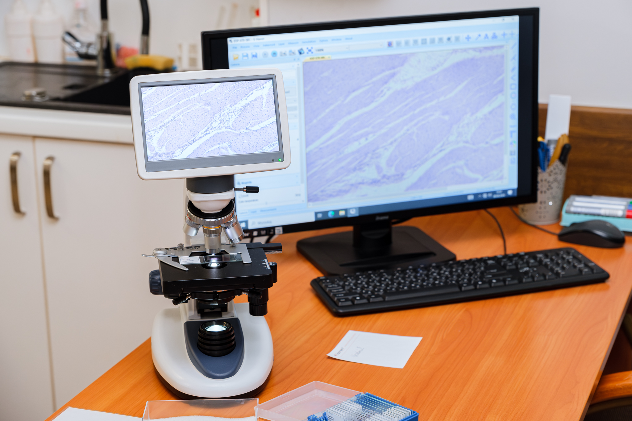
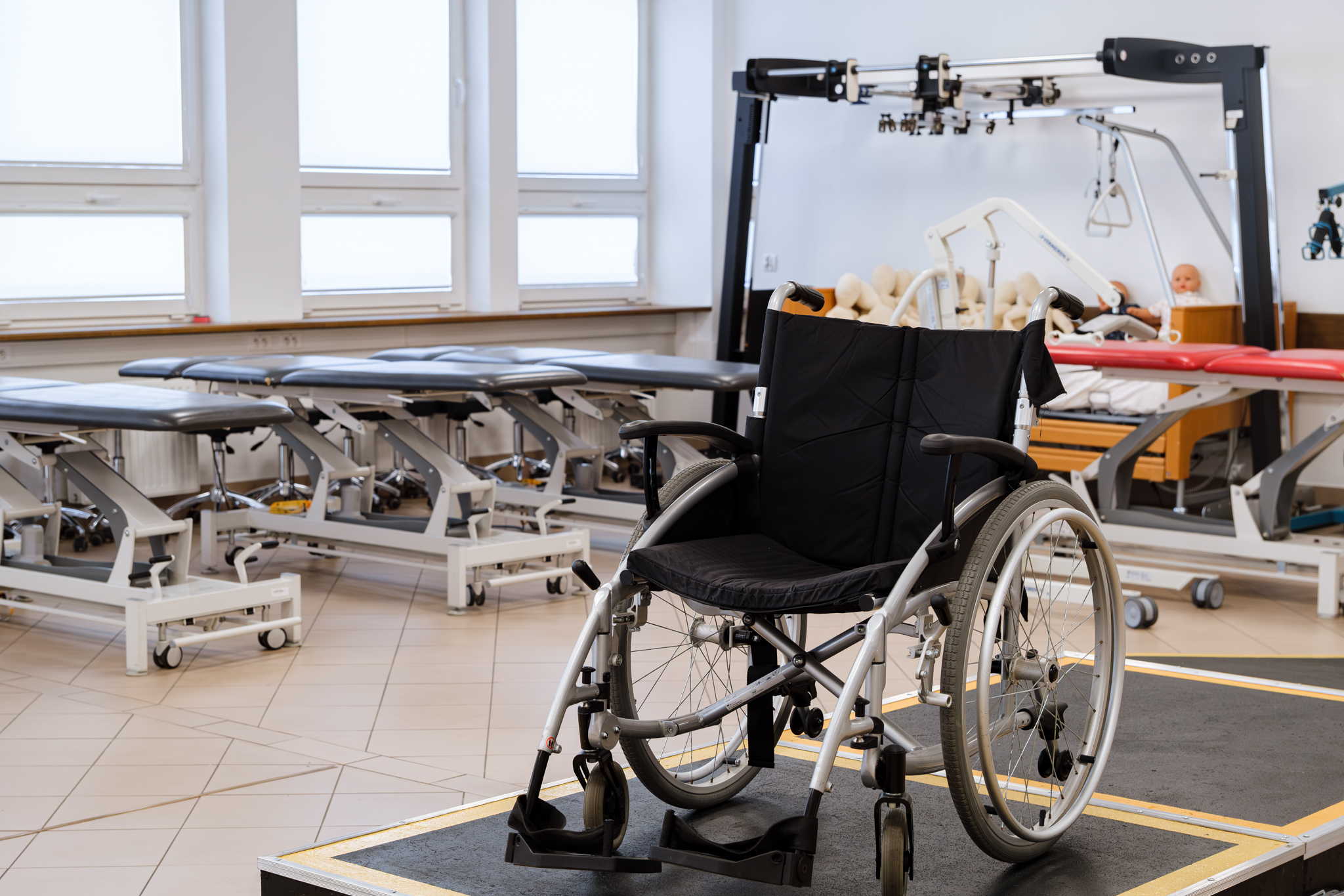
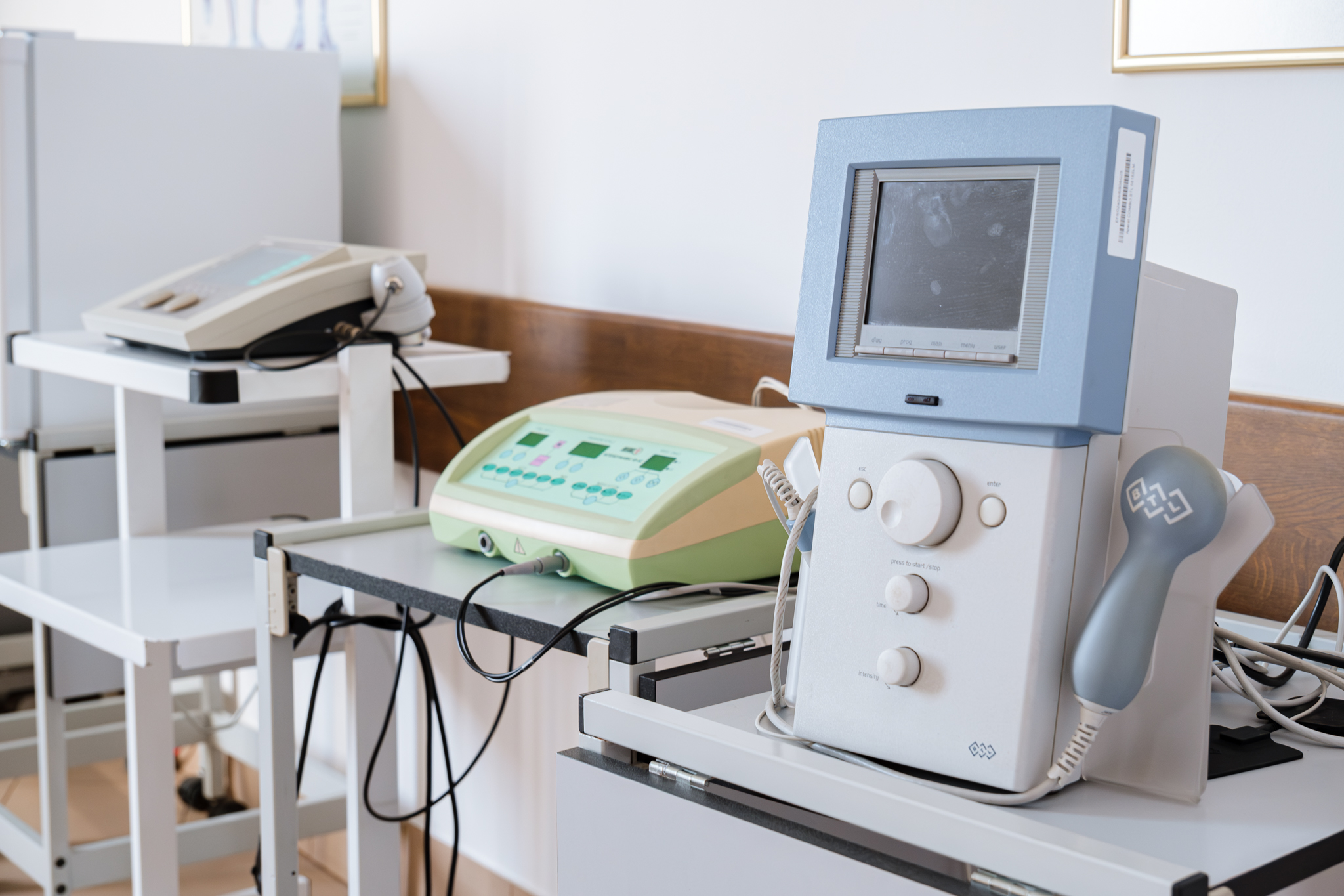
Long-Cycle Master's Degree in Medicine
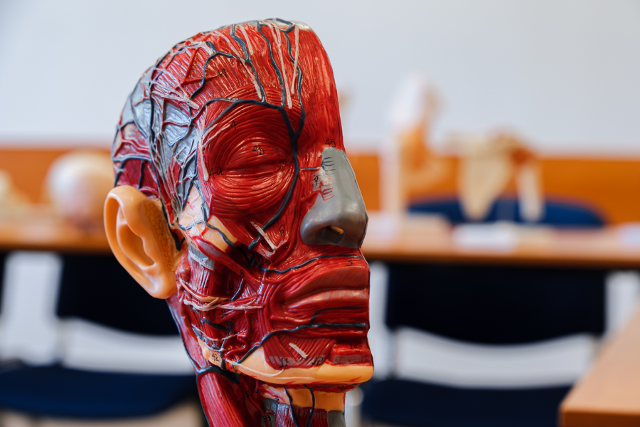
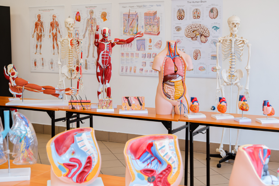
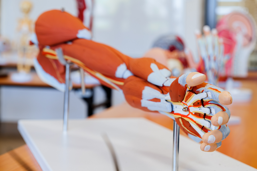
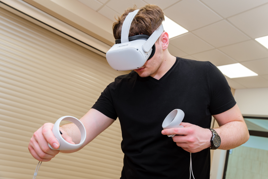
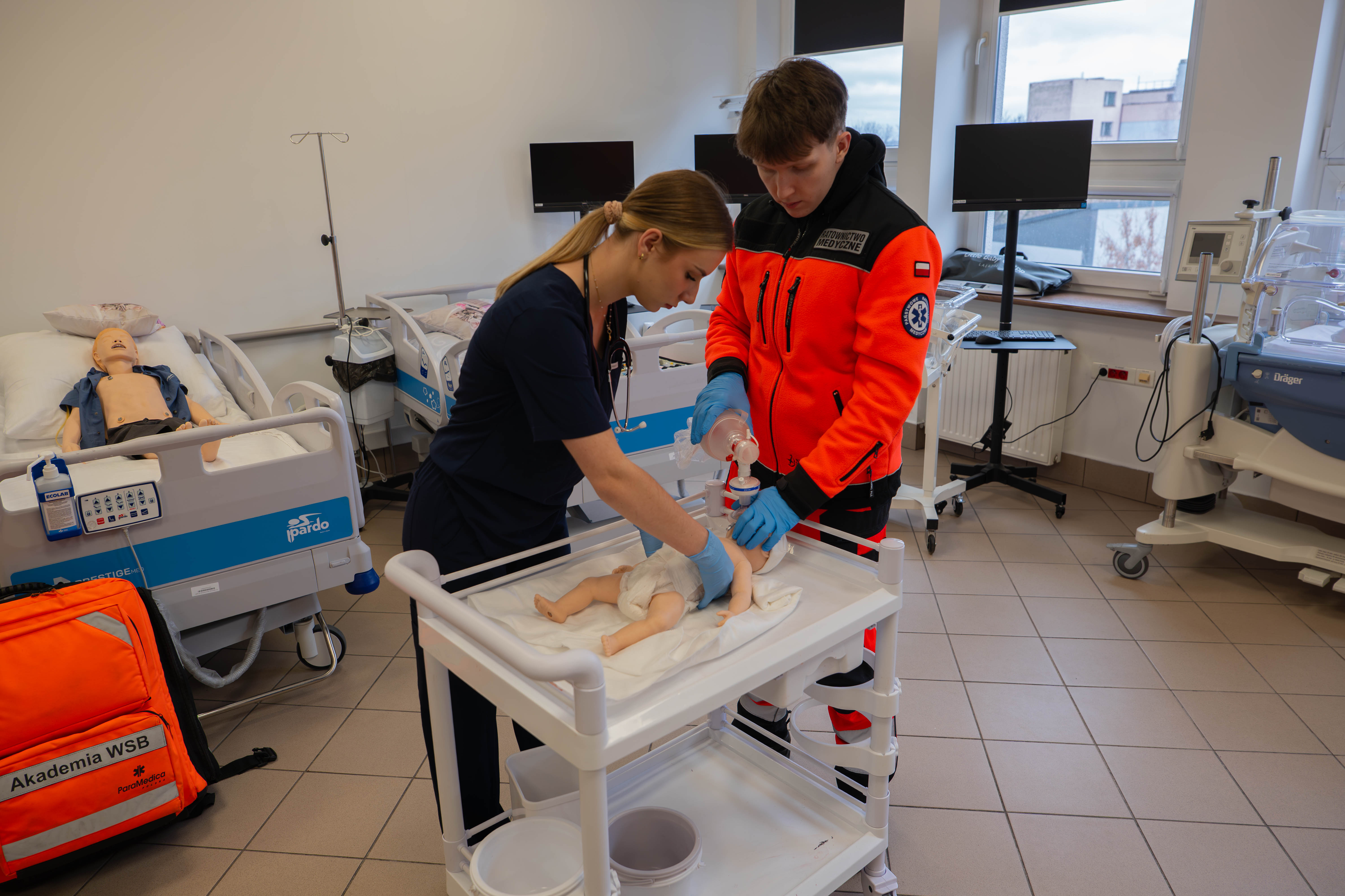
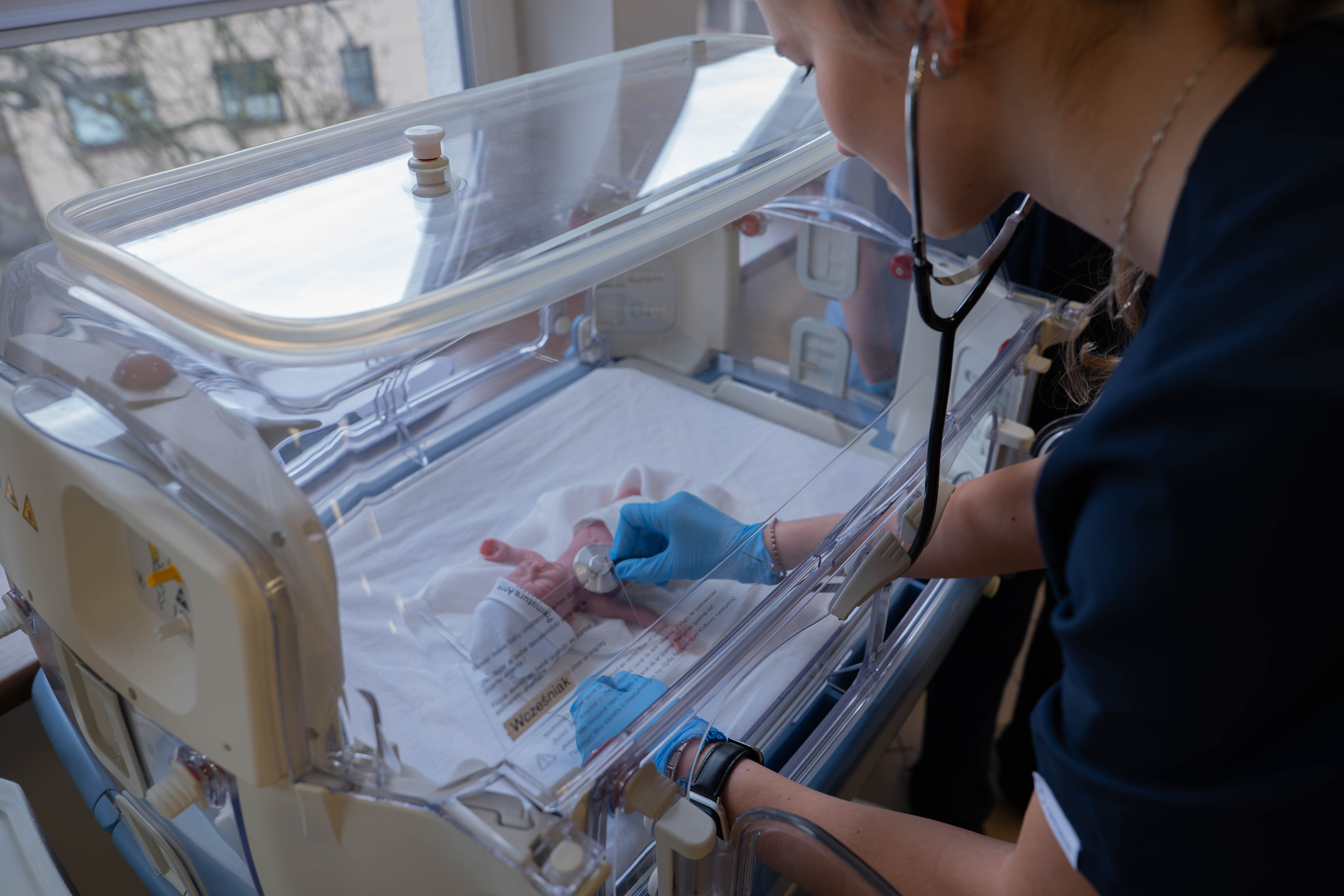
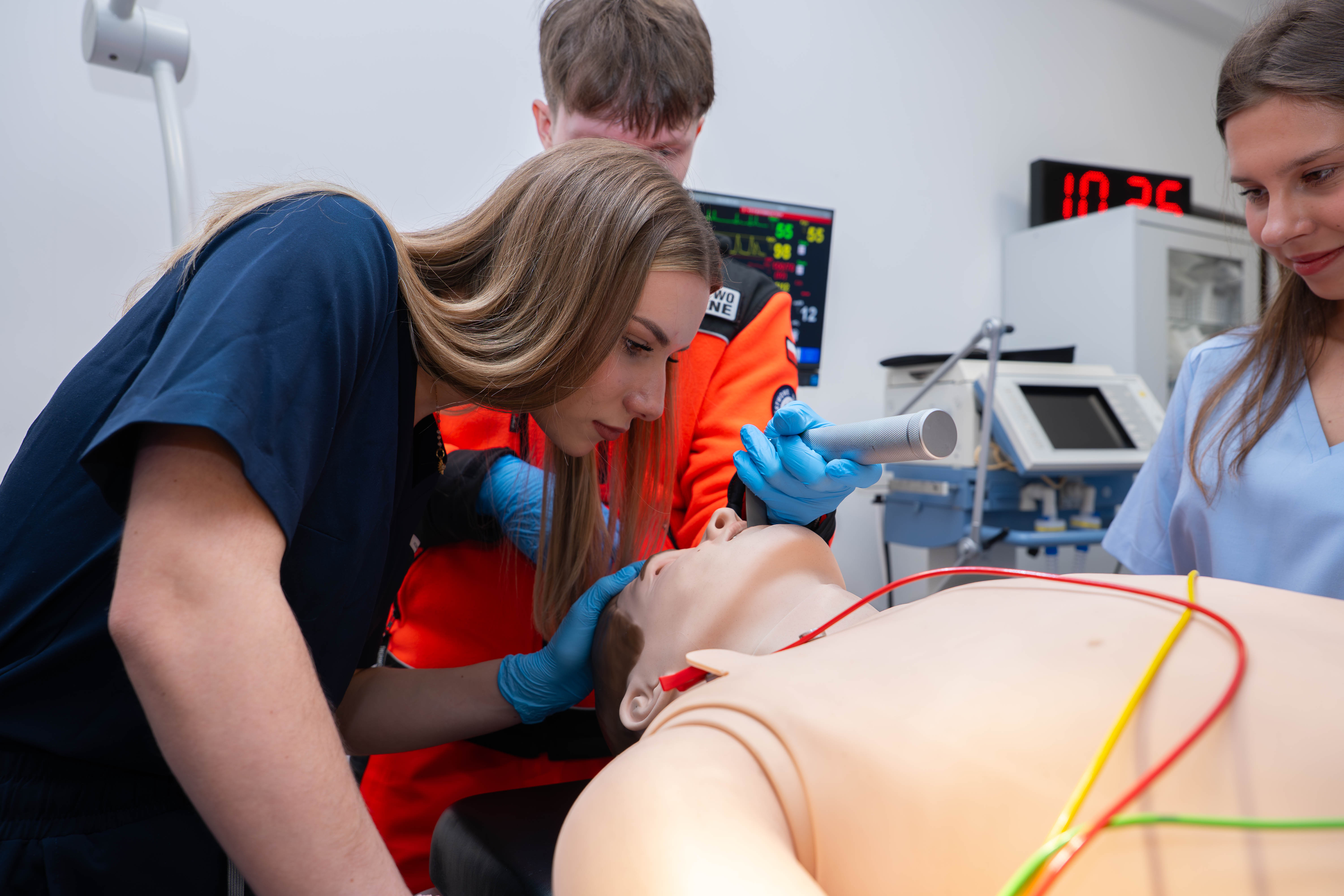
6-year full-time and part-time studies in Medicine at WSB University
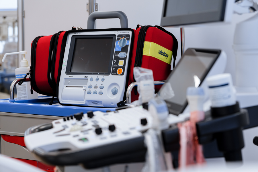
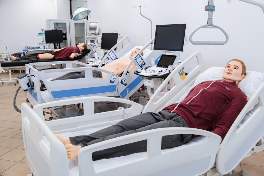
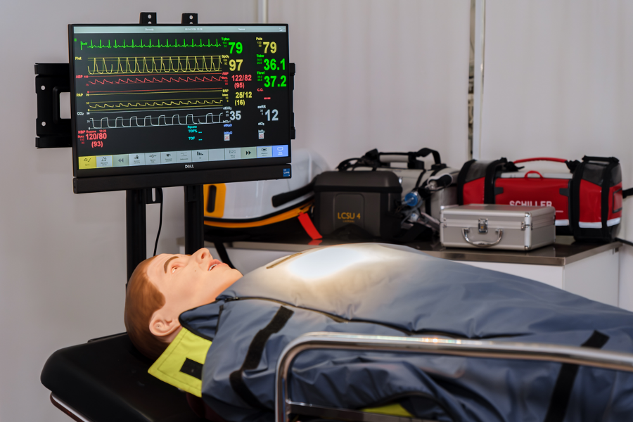
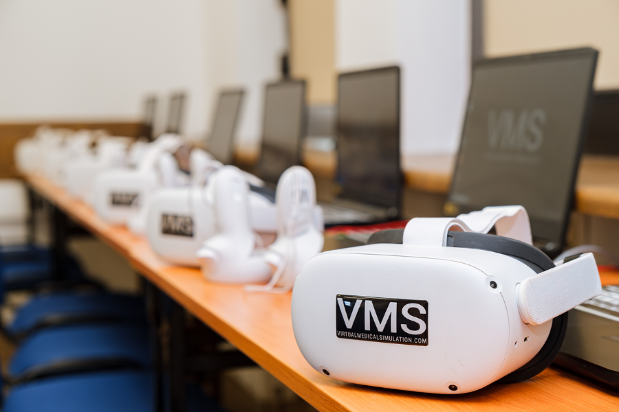
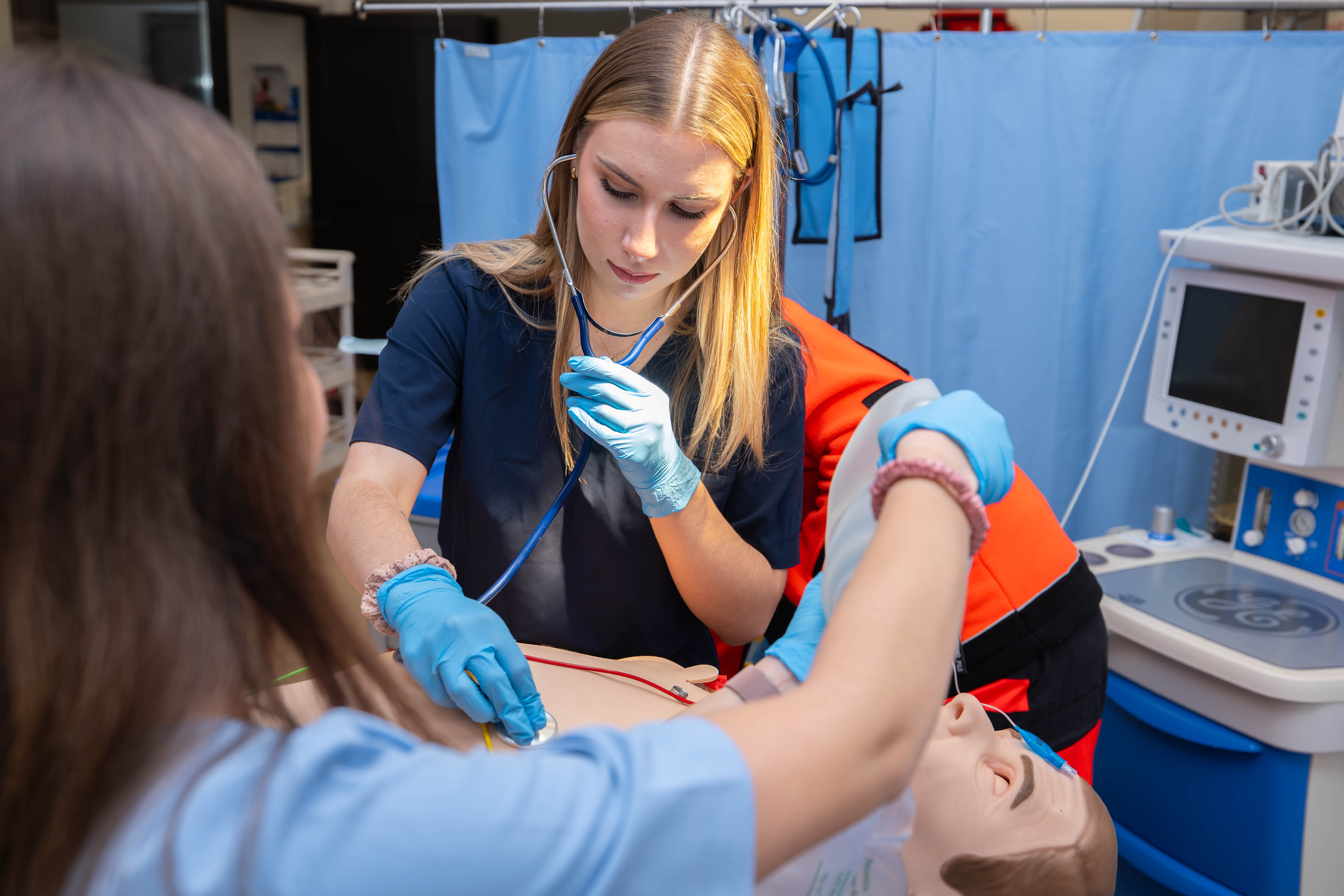
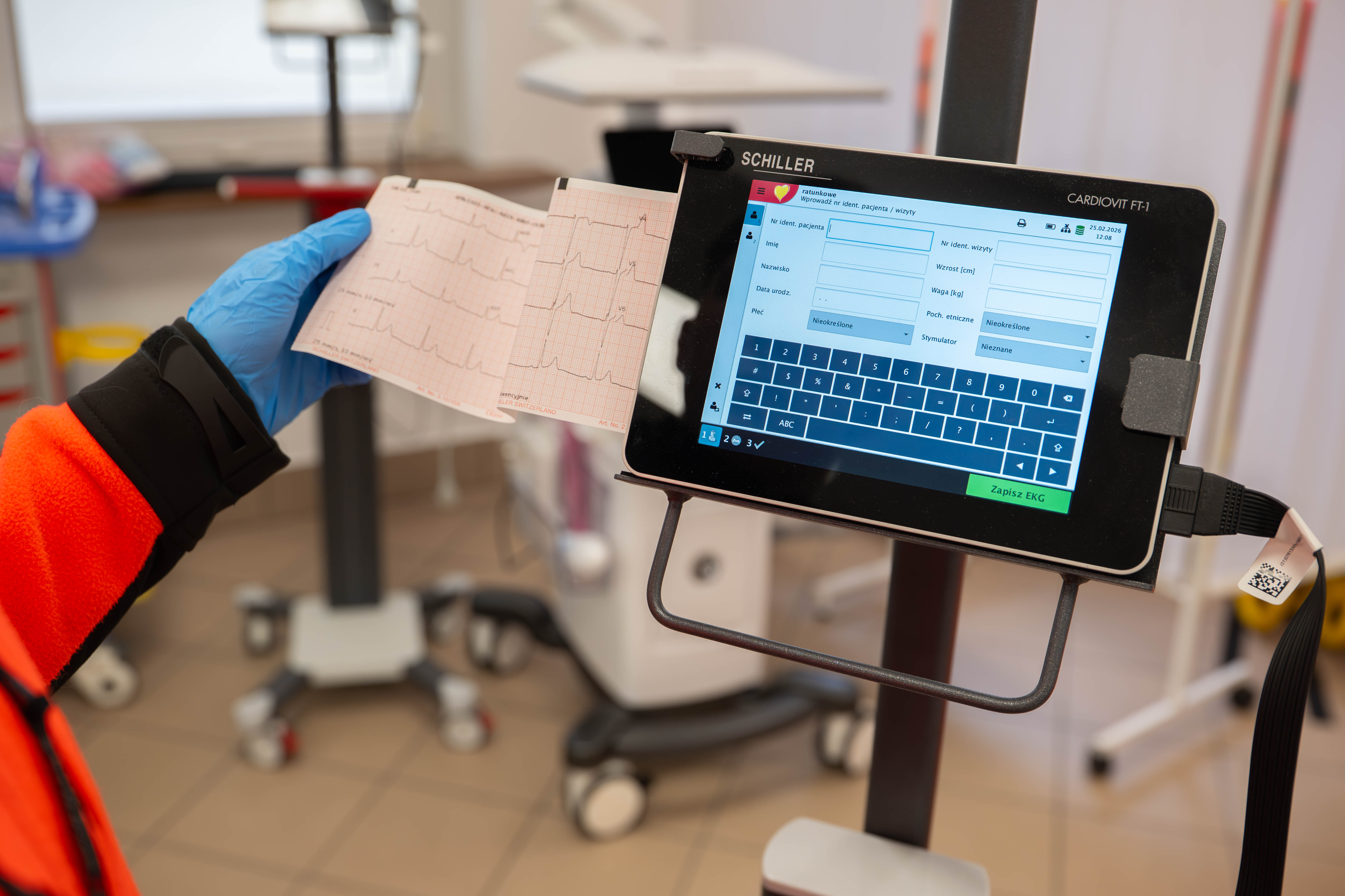
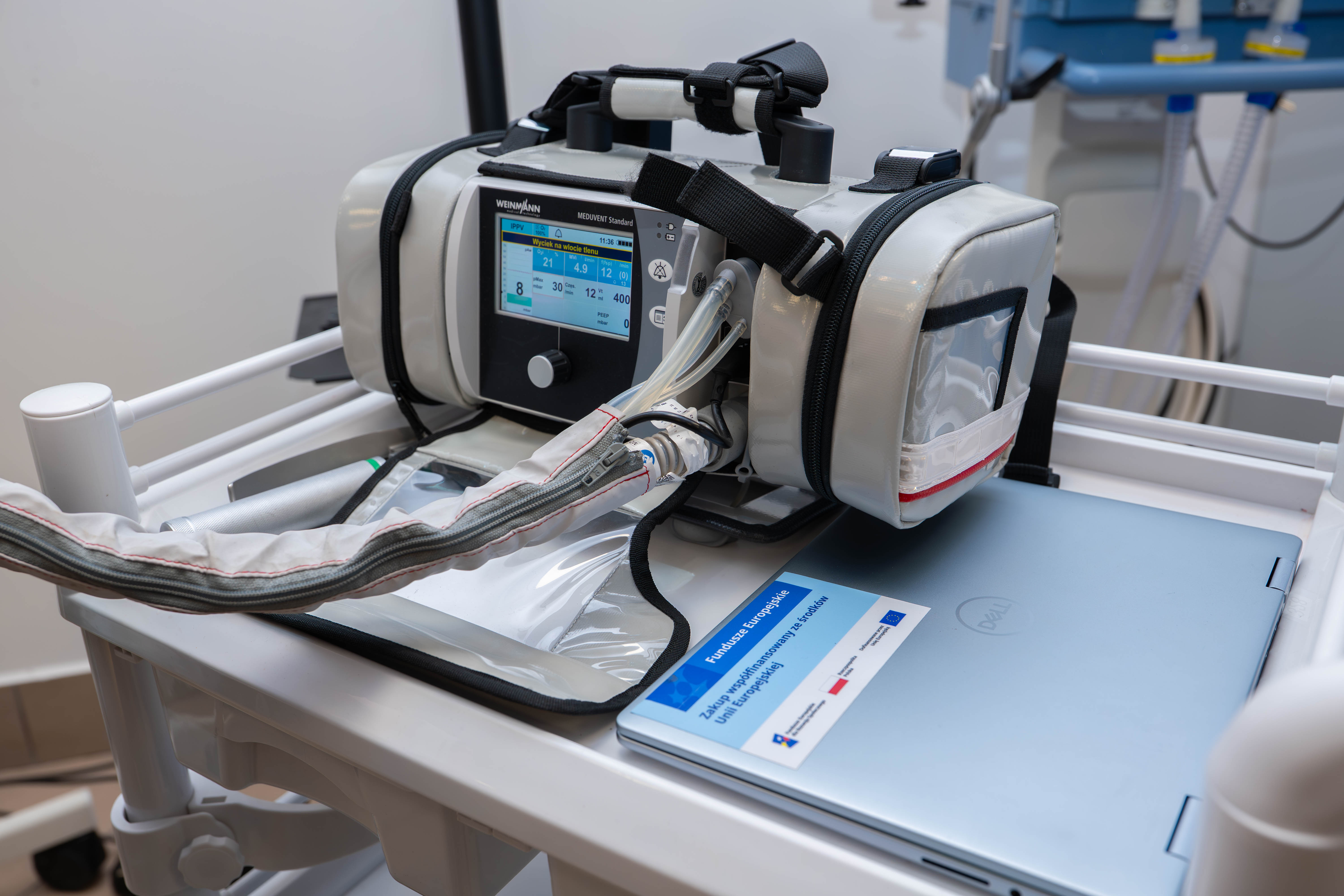
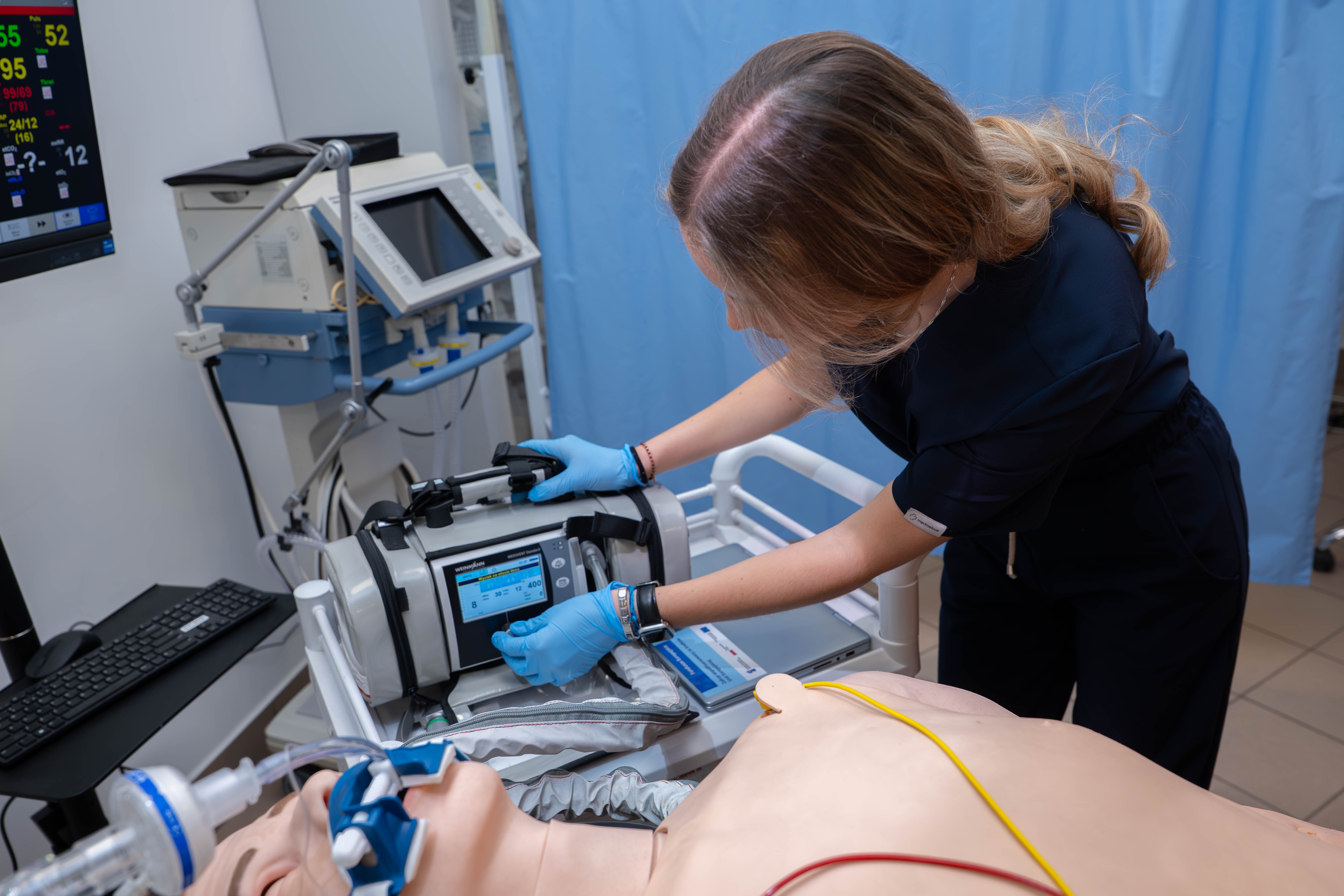
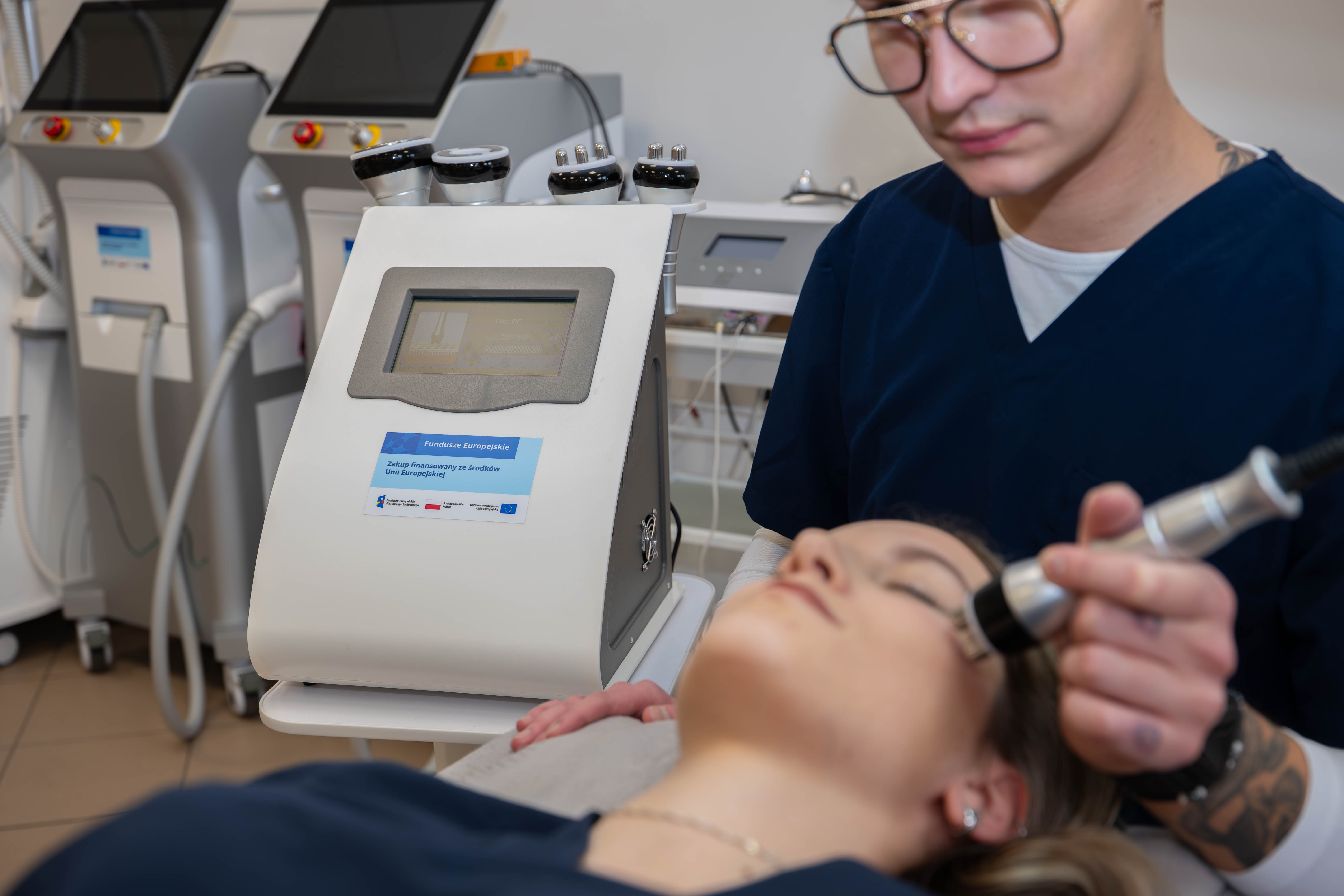
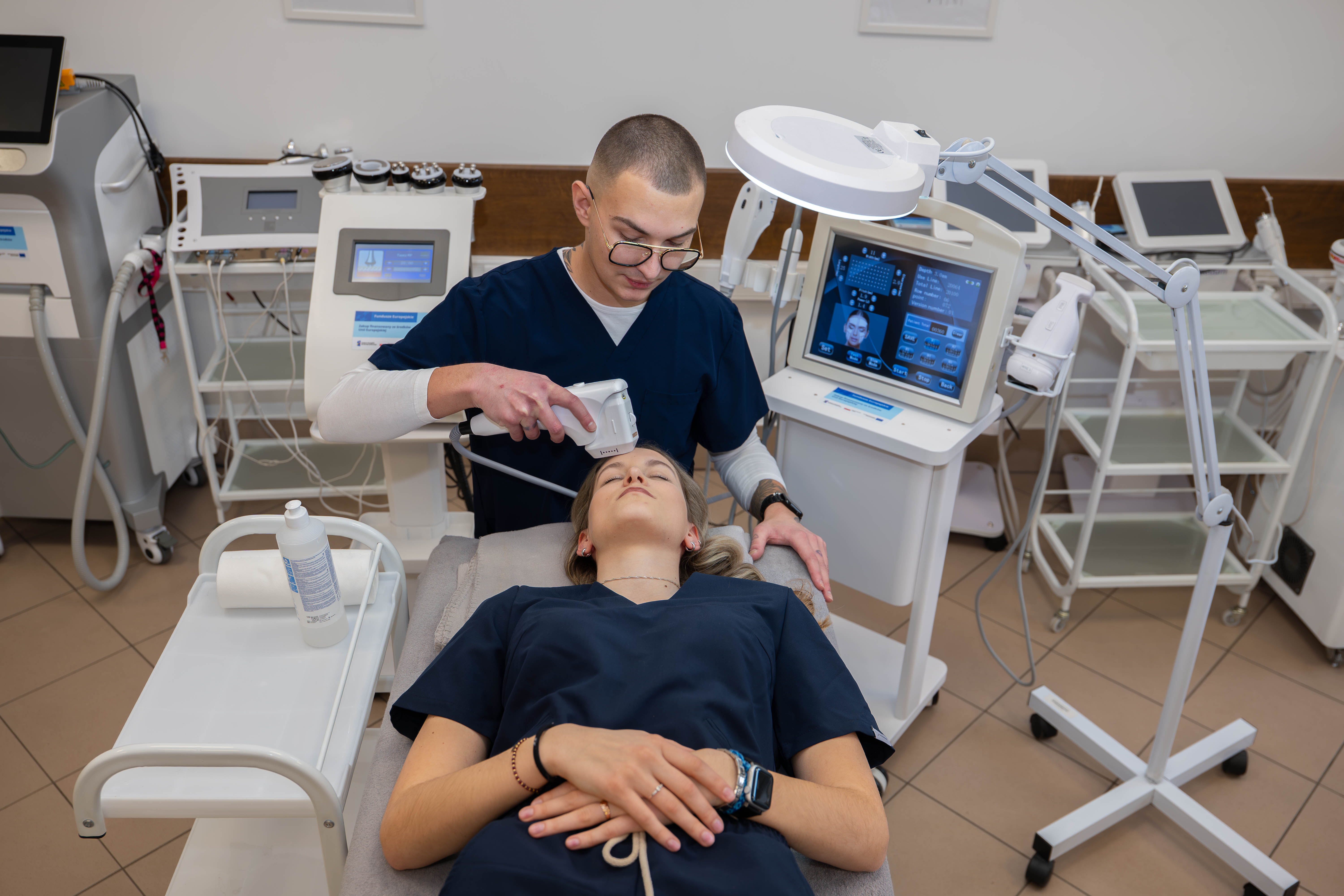
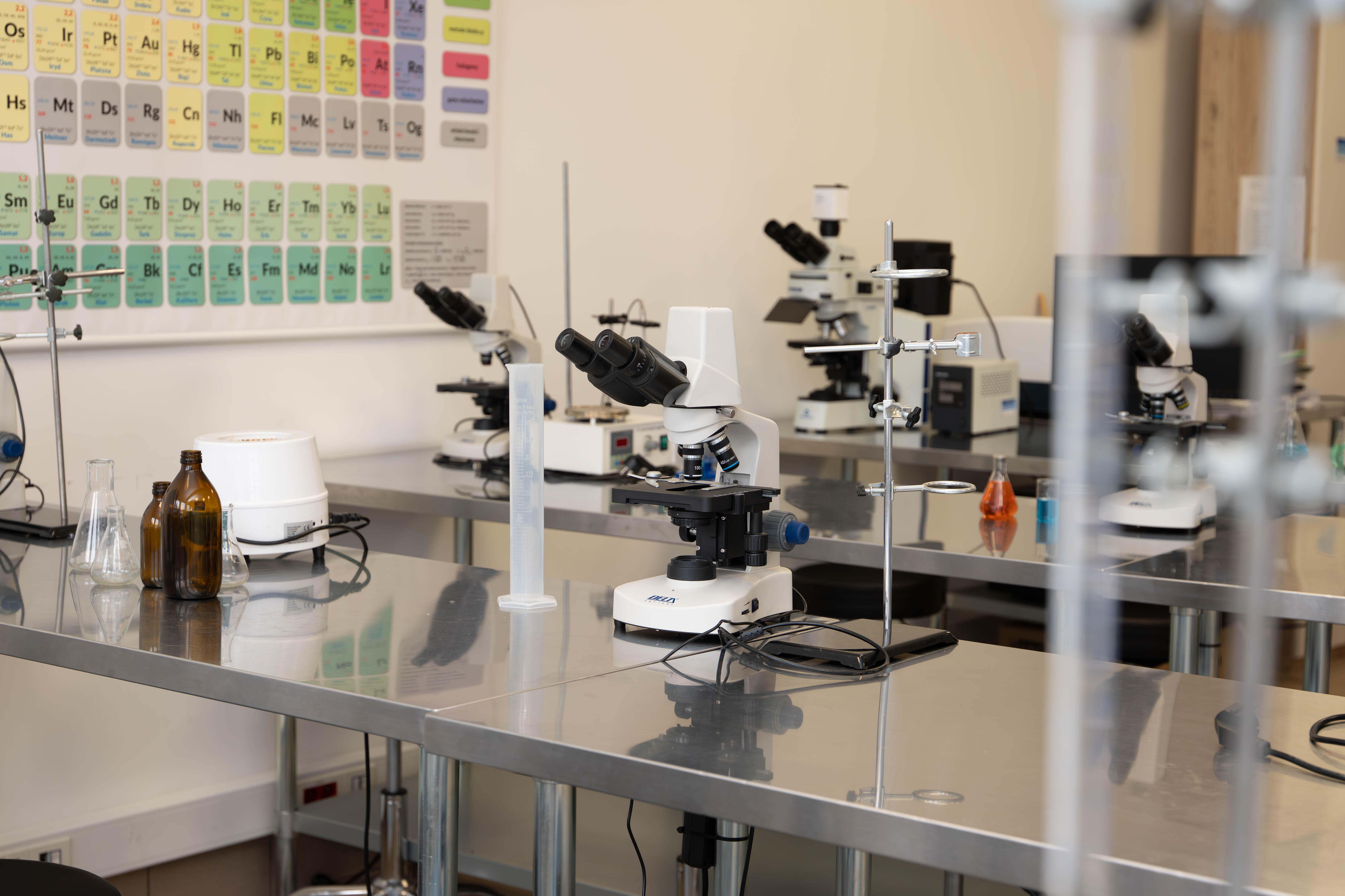
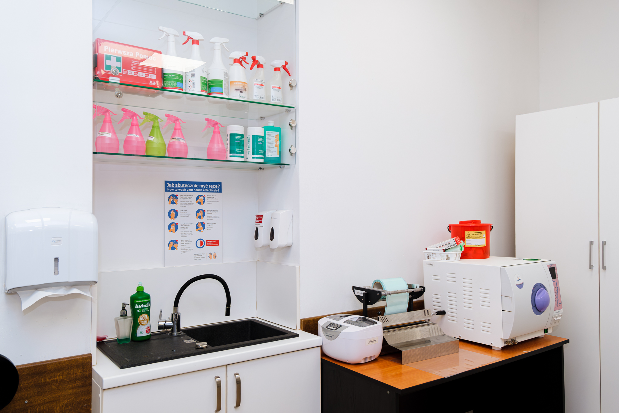
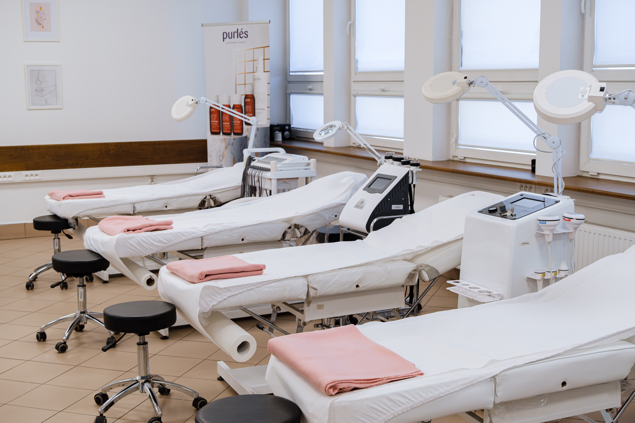
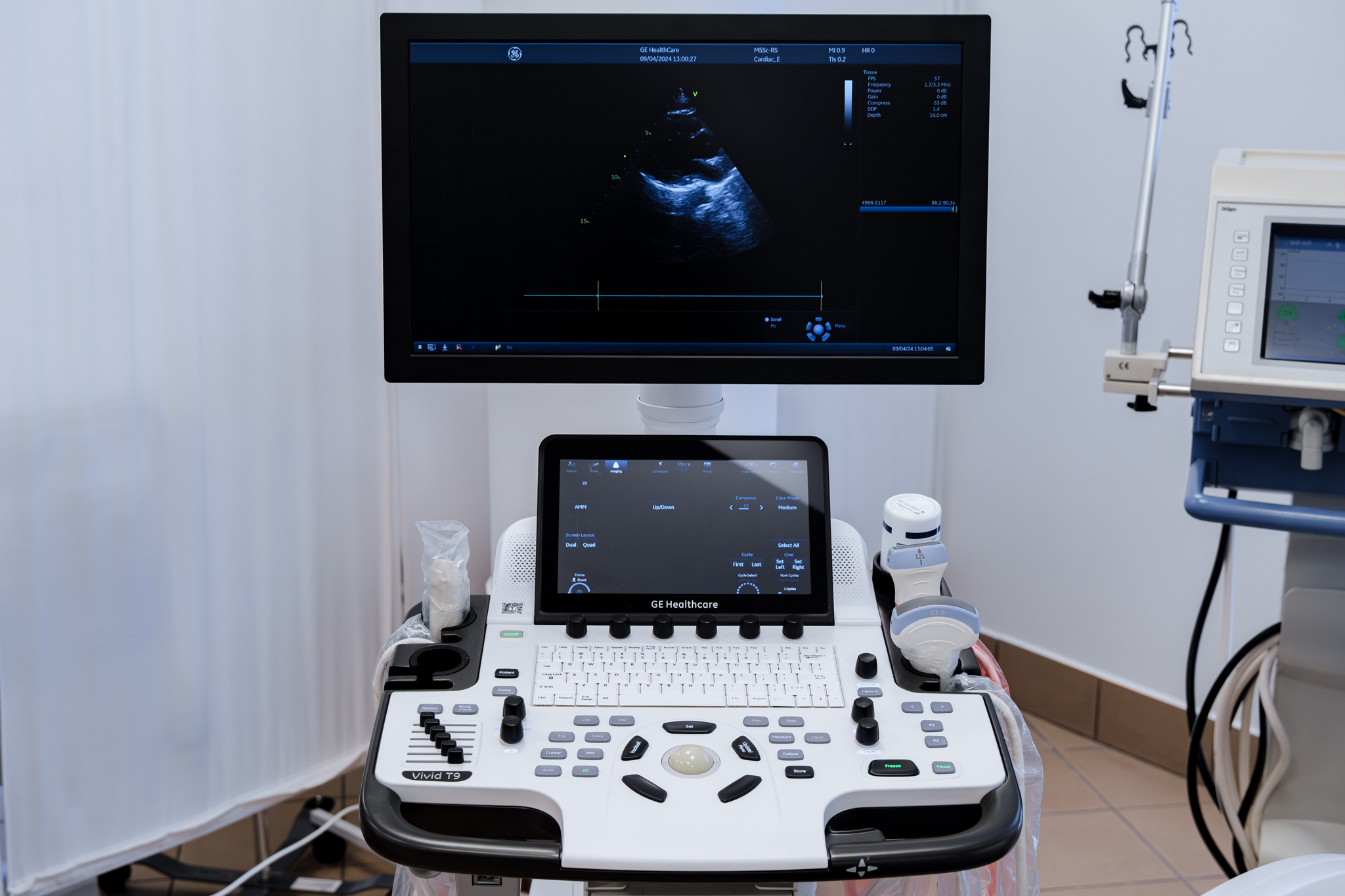
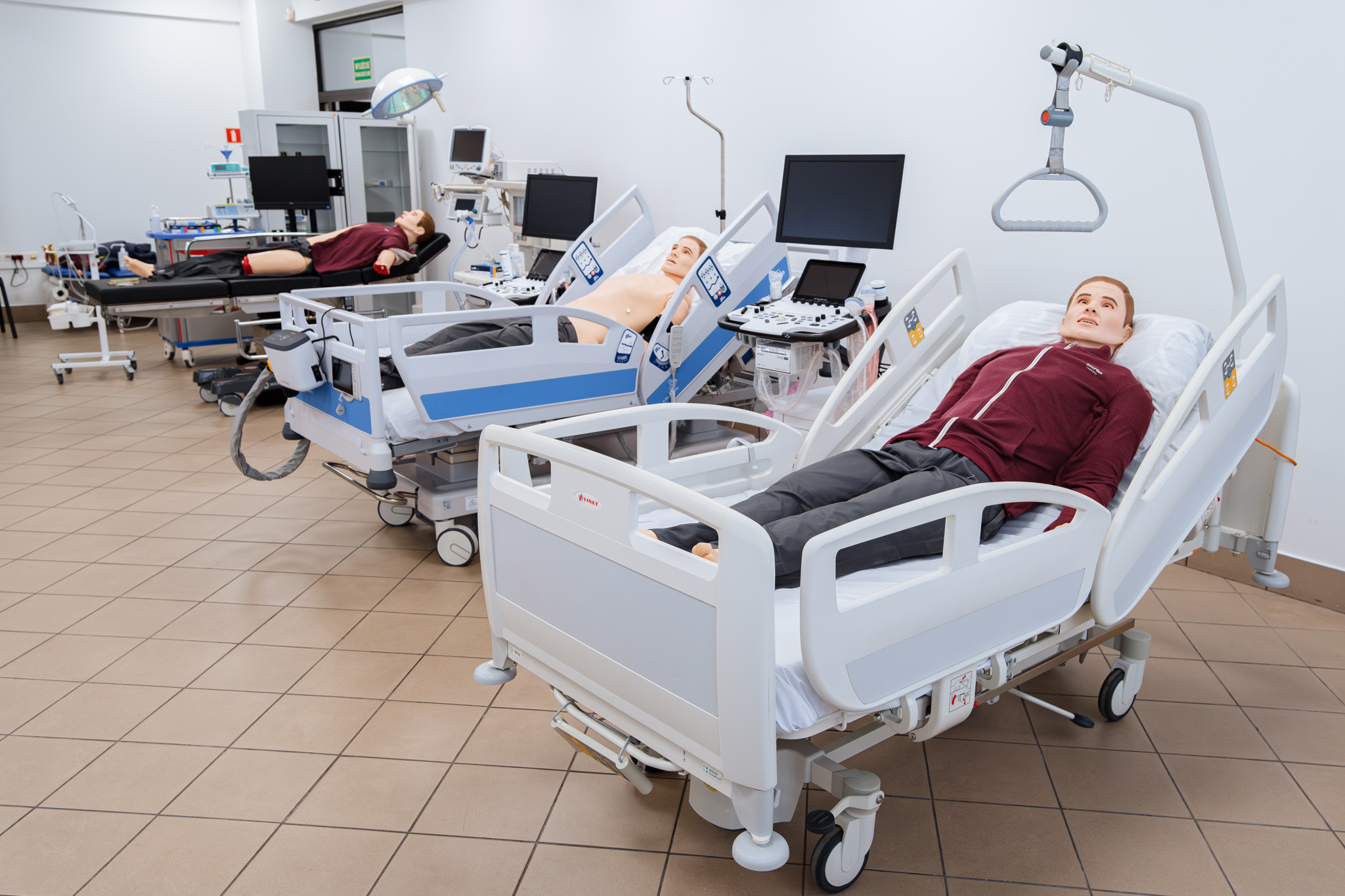
Studies in Medicine at the Collegium Medicum of WSB University primarily offer an exceptionally modern and interdisciplinary educational programme for future physicians, covering basic sciences, various aspects of medical technologies, as well as an extended clinical training programme conducted in highly specialised hospitals with a large patient flow.